Pioneer in Upper Bavaria: Chief gynecologist uses robotics in cancer surgery
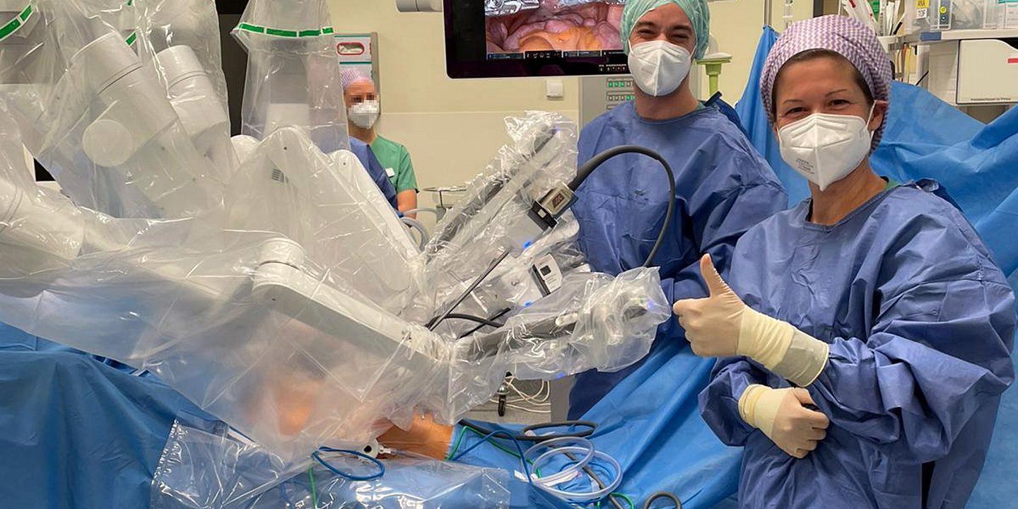
At Helios Amper-Hospital Dachau, the head physician of the Clinic for Gynecology and Obstetrics of the Upper Bavarian Hospital Association, Dr. Sabine Keim, removes lymph nodes in the area of the abdominal aorta and the female pelvis with robotic assistance. This minimally invasive surgical technique is performed using a state-of-the-art robotic system and is considered one of the latest advances in surgical gynecology.
"This is the surgical cancer treatment of the future. It is gentler and more precise than ever before,"
says Dr. med. Sabine Keim, head of gynecology at Helios Hospitals Oberbayern, as she steps out of operating room 6 at Helios Amper-Hospital in Dachau. She has just performed a so-called pelvic and paraaortic lymphonodectomy on a patient with uterine cancer, i.e. removal of the lymph nodes in the area of the abdominal aorta and the small pelvis. For the first time, the console surgeon, who specializes in robotic surgery, had a robot assist her in this challenging procedure. Just in time for International Women's Day, Dr. Keim is thus a pioneer in Upper Bavaria and Munich and is delighted about this milestone in minimally invasive surgery.
"For cancer patients, this procedure is a huge step forward," continues the focal gyneco-oncologist, who specializes with her team in the treatment of female cancers. "Sufferers can recover much more quickly." Removing the lymph nodes detects and counteracts the spread of cancer cells. Previously, this procedure was performed in an open surgery, then minimally invasive, Keim explains. "With robotic assistance, we are now technically able for the first time to achieve perfect visibility of the surgical area and maximum oncological precision and freedom of movement."
The revolutionary surgical technique is made possible by surgical robot DaVinci Xi, in addition to a special dye that prevents the unnecessary removal of lymph nodes. It ensures that Dr. Keim has almost complete freedom of movement with her instruments inside the abdomen and that the movements of her hands are precisely transmitted. The three-dimensional view of the surgical field in twelve-fold magnification enables dissection that is gentle on tissue, vessels and nerves. "We can work with very small incisions and almost without changing instruments. This shortens the operating time and contributes to a low-blood operation. The patient benefits from a faster recovery, less pain and less scarring. The likelihood of complications is reduced to an absolute minimum. Postoperative monitoring in the intensive care unit is the exception and patients can leave the hospital after a few days," explains the chief gynecologist, adding,
"We are pleased and proud to be able to offer our patients this first-class treatment."


