Breast cancer diagnosis
- Breast palpation
- Breast ultrasound (Sonography)
- Mammography (Breast X-ray)
- Magnetic resonance imaging (MRI)
- Biopsy
- Genetic testing for breast cancer
- Prophylactic breast removal
- Breast cancer diagnosis at Helios hospitals
Many women experience hormone-related breast changes and discomfort, which can also affect how the breast tissue feels during palpation. Breast tenderness or a feeling of tightness, particularly shortly before menstruation, as well as swelling or increased lumpiness during this time, are common. It is therefore important to be familiar with how your breasts normally look and feel and to pay attention to any changes. If new changes appear or a lump continues to grow, medical evaluation is recommended.
Most breast symptoms and changes are benign and not caused by cancer. Nevertheless, it is important to consult a gynecologist for further evaluation if:
-
you can feel a lump or an area of unusual firmness in the breast;
-
the breasts appear to change shape or position differently when the arms are raised;
-
a nipple becomes inverted or pulled inward;
-
changes appear in the skin of the breast or nipple;
-
there is bloody discharge from a nipple.
Breast palpation
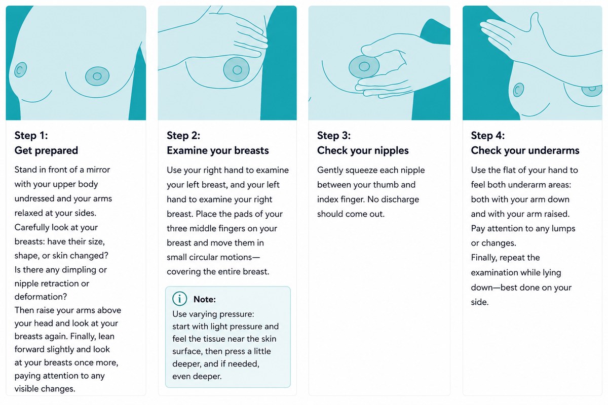
Breast self-examination can play an important role in noticing changes in the breast. Although it is not considered a screening or early detection test — a lump generally becomes palpable only after it has reached a certain size — self-examination can help women become aware of changes and seek medical evaluation when necessary.
If a suspicious lump or other abnormality is detected, the physician will first perform a physical examination. Following a clinical examination and palpation of the breasts and underarm areas, the appropriate next steps for further diagnostic evaluation can be determined.
Several diagnostic methods may be used for further assessment:
-
Breast ultrasound (sonography)
-
Mammography
-
Magnetic resonance imaging (MRI)
-
Tissue sampling (biopsy)
Breast ultrasound (Sonography)
As a rule, if a suspicious lump or other abnormality is detected during a physical examination, the gynecologist may refer the patient to a radiology practice or a specialized breast center for a breast ultrasound examination.
During breast ultrasound, high-frequency sound waves are used to create images of breast tissue on a monitor. Because different tissue structures reflect sound waves differently, ultrasound can help distinguish between solid masses and fluid-filled cysts. Since mammography and breast ultrasound visualize tissue in different ways, ultrasound is also frequently used as a complementary examination when mammography findings are unclear or require further assessment.
Breast ultrasound is often the preferred initial imaging method for women under the age of 40 who present with breast symptoms or abnormalities. Since ultrasound does not use ionizing radiation, the examination can be repeated whenever clinically necessary.
Mammography (Breast X-ray)
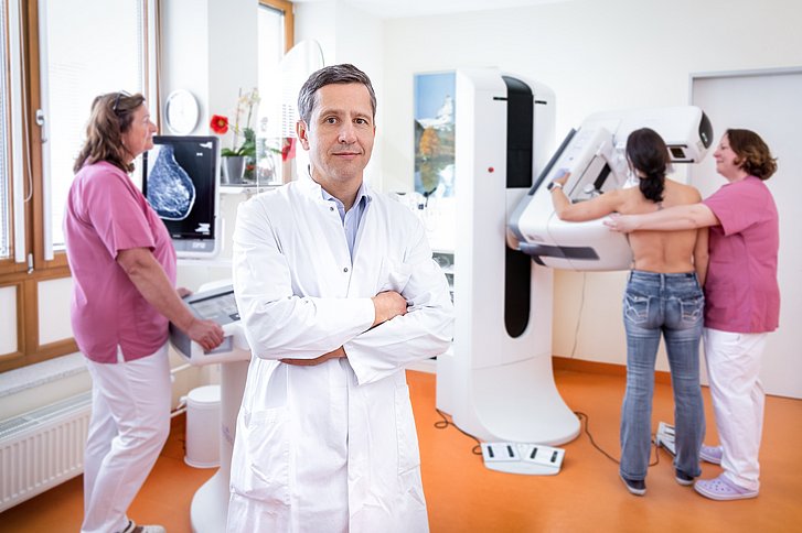
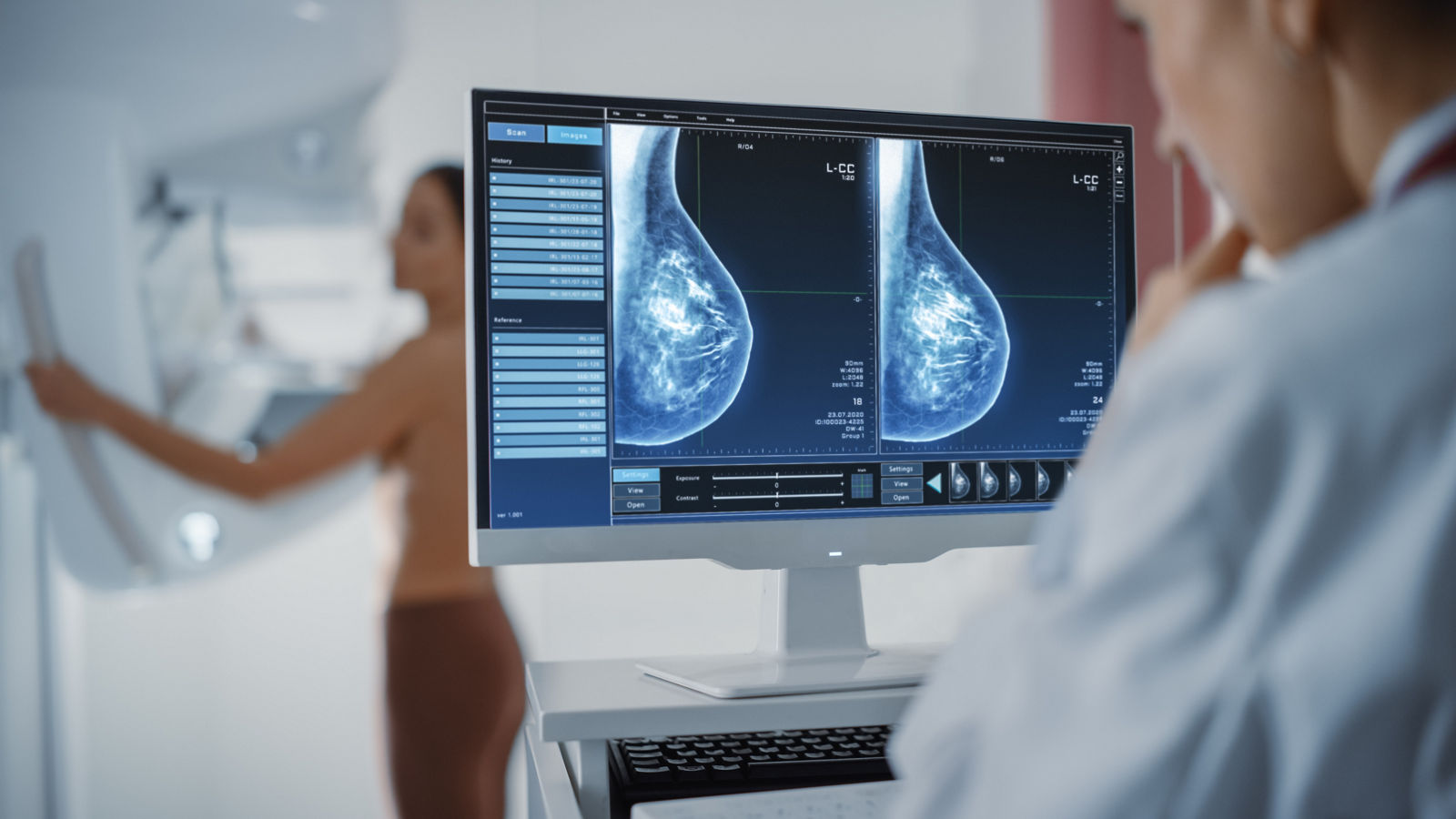
Mammography is a specialized X-ray examination of the breast. It can help determine whether a lump detected during palpation may be suspicious for breast cancer. Mammography is particularly important for detecting microcalcifications and precancerous changes. Since approximately 90% of cases of ductal carcinoma in situ (DCIS) and around 30% of invasive carcinomas are associated with microcalcifications, mammography plays an essential role in the early detection of breast cancer. It can also provide information about the location, size, and characteristics of a tumor. Changes as small as approximately three millimeters may be visualized.
During mammography, the breast is compressed between two plates, which may cause temporary discomfort. However, compression spreads the breast tissue and improves image quality, making abnormalities easier to detect while also reducing the radiation dose required. For premenopausal women, mammography may be more comfortable when scheduled during the first half of the menstrual cycle, when the breasts are generally less tender.
Mammography is typically performed using two standard views of each breast, providing images from different angles. In organized screening programs, mammograms may also be independently evaluated by two qualified specialists — a double-reading or “four-eyes” approach — to improve the reliability of the assessment.
Magnetic resonance imaging (MRI)
Breast MRI is a valuable additional diagnostic examination used to complement the findings of mammography and breast ultrasound. It is not routinely used as a screening method for all women but can be particularly useful for detecting local recurrence and for examining women with breast implants. Cross-sectional images allow doctors to clearly differentiate between scar tissue, tumor tissue, and the implant itself.
Breast MRI provides detailed images of breast tissue without the use of ionizing radiation. However, because MRI is highly sensitive, it may detect abnormalities that are subsequently found to be benign (false-positive results). For this reason, breast MRI is generally performed for specific clinical indications rather than as a standard screening examination.
Breast MRI usually requires the intravenous administration of a contrast agent, so any potential contraindications should be assessed beforehand. The examination typically takes around 20–30 minutes. Because MRI uses a powerful magnetic field, all metal objects and jewelry must be removed before the examination. Patients with pacemakers, implants, or other metallic or electronic medical devices should inform their doctor in advance so that their MRI compatibility can be confirmed.
Biopsy
A biopsy is a histological examination of tissue. If one of the preliminary examinations reveals a suspicious abnormality in the breast, a tissue sample is taken, specially stained, and examined under a microscope. Tissue analysis makes it possible to accurately determine the nature of the abnormality, the type of cells, and their growth characteristics.
There are several methods of obtaining a tissue sample, ranging from fine-needle aspiration and core needle biopsy to vacuum-assisted biopsy. Tissue is collected using a hollow needle, which is carefully inserted into the breast, usually under ultrasound guidance, and positioned at the suspicious area. A special mechanism is then used to quickly obtain a small tissue sample from the lesion.
A biopsy is performed under local anesthesia and usually does not leave any significant scars. The procedure is monitored using imaging guidance. Vacuum-assisted biopsy is commonly used to evaluate microcalcifications and is often performed under mammographic guidance. In some cases, the results of the histological examination may be available as early as the following day.
Genetic testing for breast cancer
Genetic testing for breast cancer can identify inherited changes (mutations) in genes that may be associated with an increased predisposition to developing the disease. The best-known genes are BRCA1 and BRCA2, although modern genetic panels can simultaneously analyze other genes associated with hereditary breast cancer. Testing is usually performed using a blood or saliva sample, from which DNA is extracted and analyzed in a specialized laboratory. The results are evaluated by a genetic specialist and can provide additional information about the hereditary nature of the disease, individual risks, and may also influence the choice of certain treatment options.
Genetic testing may be recommended if, on the same side of the family:
- at least three women have been diagnosed with breast cancer;
- at least two women have been diagnosed with breast cancer, with one diagnosed before the age of 51;
- at least one woman has been diagnosed with breast cancer and another woman with ovarian cancer;
- at least two women have been diagnosed with ovarian cancer;
- at least one woman has been diagnosed with both breast and ovarian cancer;
- at least one woman has been diagnosed with breast cancer at the age of 35 or younger;
- at least one woman has been diagnosed with bilateral breast cancer at the age of 50 or younger;
- at least one man has been diagnosed with breast cancer and one woman with breast or ovarian cancer.
Prophylactic breast removal
Women with confirmed inherited BRCA1 or BRCA2 mutations have a significantly increased risk of developing breast cancer as well as ovarian cancer. After such a mutation is identified, women may be offered a consultation with a specialist in hereditary cancer to assess their individual risk and discuss possible preventive measures. In addition to more intensive surveillance and regular screening, one option may be a prophylactic bilateral mastectomy — the removal of breast tissue from both breasts before cancer develops.
A prophylactic mastectomy can significantly reduce the risk of developing breast cancer, although it cannot eliminate the risk completely. In many cases, the procedure can be performed while preserving the breast skin and, when appropriate, the nipple and areola, with immediate breast reconstruction using implants or the patient’s own tissue. The decision to undergo this procedure is made individually, taking into account the specific genetic mutation, age, family history, overall health, and the patient’s personal preferences.
BRCA1/2 mutations are also associated with an increased risk of ovarian and fallopian tube cancer. For this reason, after family planning has been completed and at an appropriate age, preventive removal of the fallopian tubes and ovaries (risk-reducing salpingo-oophorectomy) may also be considered. The optimal timing of this procedure depends, among other factors, on whether a BRCA1 or BRCA2 mutation has been identified. Decisions regarding preventive strategies should be made after detailed consultation with a genetic specialist, gynecologist, and breast specialist.
Breast cancer diagnosis at Helios hospitals
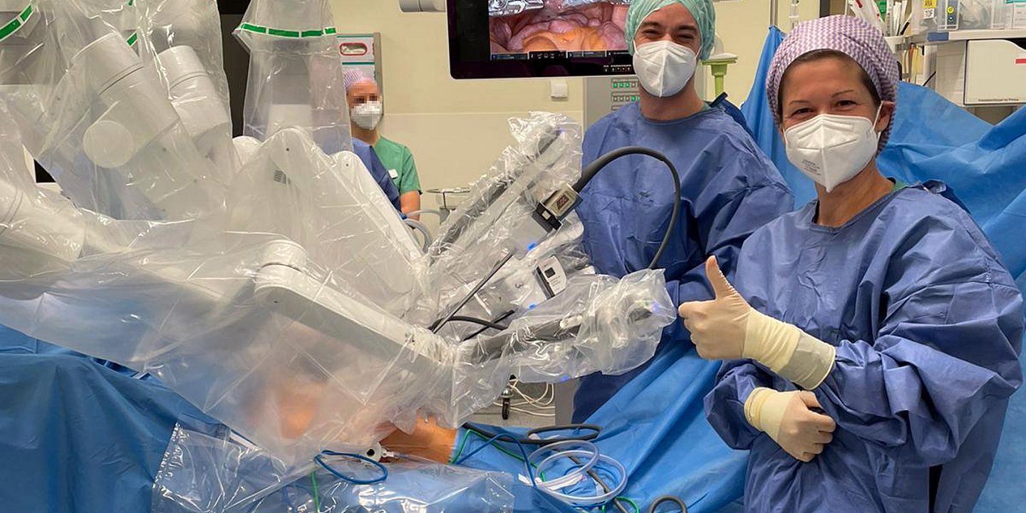
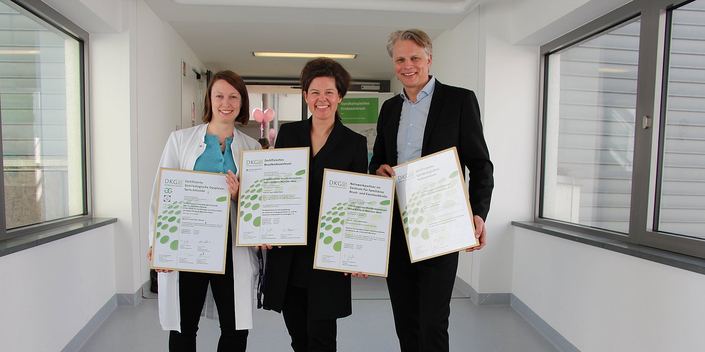
At specialized Helios breast centers, breast cancer diagnosis is performed by experienced specialists using modern, high-precision medical equipment and in accordance with current medical standards.
To determine a targeted and individualized treatment strategy, Helios specialists discuss the diagnostic findings of breast cancer patients at multidisciplinary tumor boards. These meetings bring together all specialists involved in the patient’s care — from diagnostic experts and gynecological surgeons to pathologists, medical oncologists, and radiation oncologists — to determine the most appropriate treatment plan.
The combined expertise of Helios specialists ensures that the different aspects of the disease are taken into account and that treatment is based on the latest medical knowledge. Helios physicians also participate in specialized medical societies, scientific conferences, and research activities both in Germany and internationally.
If you are interested in arranging diagnosis or treatment at one of the Helios hospitals, you can submit a request using the contact form on our website or contact us via the email address provided at the top of the website.
Learn more about arranging treatment at Helios hospitals ⟶
Do you need more information about Helios Hospitals or want to schedule your treatment?